ADA standard
The digital studio of the Dr Lauc Dental Imaging in Ilica 19 is standardized according to the American Dental Association (ADA) SCDI TR 1023 standard.
Doc.dr.sc. Tomislav Lauc is member of the Central Committee and the Board of specialization of European Academy for DentoMaxilloFacial Radiology. The protocols used by Dr. Lauc Dental Imaging are taken from the protocol of the European Academy.
Application of personal protective equipment
The equipment meets all the regulations for radiation safety. X-ray radiation is filtered through filters of a satisfactory and regulated thickness (2.5 mm AI). There is a protective apron for patients, whose protection is equivalent to 0.3 mm of lead, of satisfactory protective power. Apart from the apron. there is an additional thyroid shield with a protective power of 0.5 mm of lead.
A report of the Ruđer Bošković Institute on the testing of diagnostic radiographic equipment in Dr Lauc Dental Imaging in Ilica 19:
The advances in radiographic technology have enabled the highest level of security in today’s radiographic diagnostics in dental medicine. Contemporary devices are safe and apply very low levels of radiation. There is no dispersion of x-rays during imaging. Each piece of equipment is certified on the State Office for Radiation Protection, Croatia (SORP) by the Ruđer Bošković Institute (IRB) and the
SORP. Regular controls and certificate renewals are conducted for all equipment according to the Act on Ionizing Radiation Protection and Safety of Ionizing Radiation Sources, Ordinance on the Register of Activities, Requirements and the Manner of Issuing, and the Validity of Licenses for Work with Sources of Ionizing Radiation and the Use of Sources of Ionizing Radiation, the State Plan and Program of Ionizing Radiation Safety Measures and Emergency Interventions, the Ordinance on Ionizing Radiation Conditions and Safety Measures for Performing Activities with Radiographic Devices, Accelerators and Other Devices that Produce Ionizing Radiation, the Ordinance on Ionizing Radiation Conditions and Safety Measures for Performing Activities with Radioactive Sources, the Ordinance on the Conditions for Applying Ionizing Radiation Sources in Medicine and Dental Medicine, the Ordinance on the Conditions, Deadlines and Ways of Acquiring the Necessary Professional Education and Renewing Knowledge on the Application of Ionizing Radiation Safety Measures, and the Ordinance on Health Conditions for Exposed Employees, the Frequency of Examinations and the Content, Manner and Deadlines of Storing the Data on the Mentioned Examinations.
The imaging room is isolated by barite mortar and the doors are covered with a protective lead foil. The staff handling the radiographic devices is composed of medical radiology technicians with a state exam and a license to work with radiographic devices who have undergone professional education for the application of all safety procedures.
The type of radiographic films is an important factor in determining radiographic exposition. In 1993, the Nationwide Evaluation of X-ray Trends (NEXT) program classified intraoral radiographic films into three categories: D (slowest), E and F-speed (fastest). The greater the speed of the film is, the lower the need for radiation exposition. For example, the radiation dose for D films is 1.7 mGy per film, for E films 1.3 mGy and for F films 1 mGy. A change of films from D to E reduces exposition 30-40 %, from E to F 20-25 %, and from D to F an astounding 60 %.
Digital technology largely reduces the need for radiographic radiation. As exposition is significantly reduced, patient exposure during imaging is reduced to a minimum. Specialized programs with additionally lowered levels of radiation are used for children. Patient protection is the primary responsibility of each of our medical radiology technicians and it is given our full attention.
Radiation protection in pediatric radiology
Image Gently Alliance
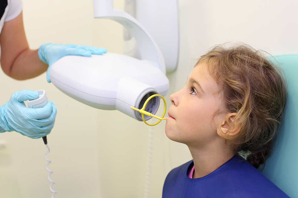
It is compulsory in planning prosthetic therapy in orthodontics and in planning implant position or extracting unbudded teeth in oral surgery. A panoramic image can be made for children as well, as it uses an adjusted, decreased level of radiation for children.
Radiologists play a key role in health services for children. Radiology and CT are critical in diagnosing diseases in children and they have an effect both on therapy and improving its outcomes. The responsibilities of radiologists, as well as all team members, is the safety of every child during a radiological examination, which needs to be customised to suit the child.
Research has shown that children are more sensitive to radiation and that they can develop negative changes in their bodies after receiving increased doses of radiation. This is why we give children special attention. We exclusively use radiographic systems designed to account for working with children, which includes a drastic decrease in dosage and imaging time.
We are the first in Croatian dental medicine to have joined the Image Gently project, where we promote caution in making decisions on imaging a small patient among our colleagues.