3D imaging

3D diagnostics, CBCT fields and CBCT viewing

3D diagnostics

Dr Lauc Dental Imaging in Ilica 19 is the first in Zagreb to enable using a 3D CT based on a cone beam (Cone Beam CT); a dental image of high quality necessary for making a diagnosis and planning dental procedures. High-quality procedures are performed safely and effectively.
SCANORA 3D combines low levels of radiation with fast image synthesis and high precision. A standard resolution produces the lowest level of radiation, while a high resolution increases precision and the quantity of information on the image. The smallest achievable voxel (volume element – the unit of rendering size) has a size of 0.13 mm on a 3D S field. To decrease the amount of radiation the patient receives, the resolution is decreased with the increase in the size of the field.

Reduced need for radiation

The level of radiation with all imaging fields is low. Radiation on an S 6×6 cm is comparable to 2 digital panoramic radiographs, while capturing a larger field in high resolution to several panoramic expositions. Creating an image by means of x-rays is a compromise between image quality and radiation quantity. By using the SCANORA 3D device this dilemma is successfully resolved by combining a high image quality with a large quantity of information and low levels of radiation. The key factors are the production of pulsating x-rays of a high technological degree and the latest ART method of image reconstruction.
SCANORA 3D enables a precise decrease of radiation levels depending on the necessary basic diagnosis or healing follow-up.

High diagnostic value

The SCANORA 3D system enables a new and innovative introduction into dentomaxillofacial anatomy and the performance of diagnostic tasks. It is very easy to choose the best 3D technique for a specific problem before moving on to plan an appropriate treatment and follow up on the results. The user can choose which system characteristics to use, which results in an excellent diagnostic performance.

Outstanding mathematical 3D image synthesis

For the first time in dental radiology, SCANORA 3D uses an advanced algebraic reconstruction technique (ART) to reconstruct spatial 3D data. ART enhances quality and is less susceptible to the main causes of image imperfection (moving, reconstruction and metals). The ART method requires a smaller amount of radiation than usual algorithms, the image quality is stable and the level of achieving a quality image is very high.

Excellent possibilities of image management

The system uses a wide array of diagnostic tools alongside all the functions of standard image processing. Among others, it has the function of showing the surface area and volume with a section of the anatomical plane/height, which is used to virtually remove obtrusive structures and create a good view of the area to be studied. Other options permit increasing the visibility of the tooth, bone and soft tissue. Also, it is possible to regulate the color view and the levels of shading and transparency.

Simple to use

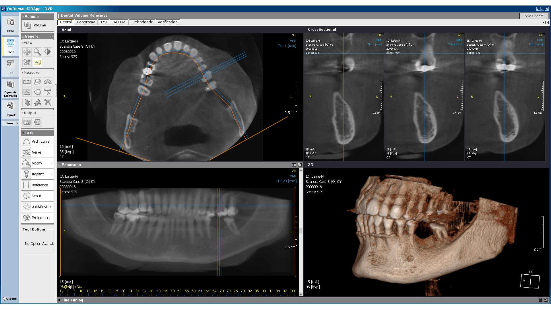
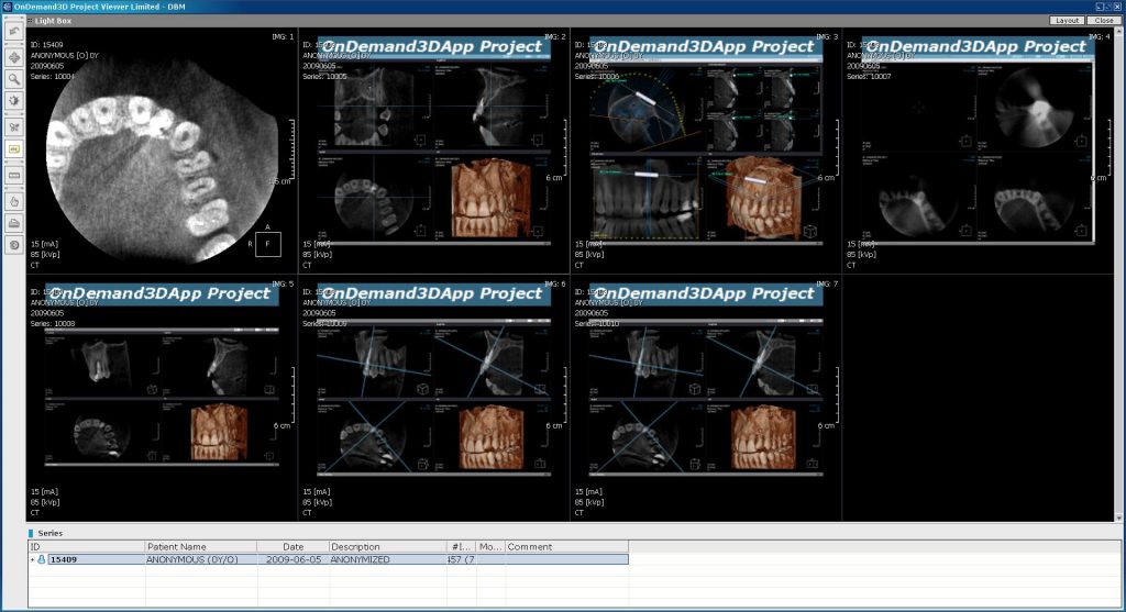
Clinical cases are shown on CDs and can be viewed directly by means of an accompanying viewer. For faster loading, we recommend copying the CD content onto the computer’s hard drive and loading it from the computer. If the doctor of dental medicine so wishes, the images can be printed on a larger (35×43 cm) or smaller film dimensions (25×30 cm). Alongside the standard 3D visualization computer program that comes with the image, most other programs supporting the DICOM format can be used. That way the system is available and open to many adjustments and changes, and it can be upgraded using the leading concepts of implantology.

CBCT fields of view

A 3D Cone Beam based CT (Cone Beam CT) offers a wide variety of viewing combinations.

3D S field

A small field (6×6 cm) is meant for planning procedures in one sextant or quadrant. A section of the antagonistic quadrant.

Indications:
Sextants: lateral teeth (3-8) of each quadrant or upper and lower front teeth.

3D M field

A medium field (7.5-10 cm) is suitable for viewing the entire dental arch, including the mandibular canal. This field is also used to create an implant matrix in guided implantology.

Indications:
Upper and lower dental arch
Upper dental arch and sinuses
Lower dental arch and mandibular canal

3D L/XL field

Large fields (7.5×14.5 cm / 10×14.5 cm) are ideal in situations where both jaws need to be examined. The image shows the TM joints and the upper section of the cervical spine. The choice between these two fields is made by radiology technicians.

Indications:
Maxilla and mandible

Unlimited number of viewing combinations

A single 3D image can be a source for any number of high quality cross-sections: panoramic, axial, transversal, diagonal, sagittal, inclined and three-dimensional, and they can all be made with just a few mouse clicks. The thickness of each image, i.e. cross-section, can be adjusted to fit the viewer’s needs. This way you are able to view any combination of cross-sections on your screen. With every set of images we will set up several “projects”, that is combinations of cross-sections that can be viewed.

Panoramic and teleradiographic images

Panoramic and teleradiographic images can be produced by reconstructing the 3D volume of the 3D L/XL field individually with their own option to regulate layer thickness.

Diagonal cross-sections

It is easy and quick to produce a sequence of diagonal cross-sections for any element of maxillofacial anatomy.

Axial images

Viewing a series of axial images is an excellent way to rate the status of the sinuses, air ways, sediments in ducts, and the position of third molars and impacted teeth.

Transversal images

A transversal view provides a clear picture of the sinuses and the positions of the roots in the bone and in relation to other structures.

MPR (MultiPlanar Reconstruction)

A multidimensional reconstruction is a basic way of visualizing anatomy. Sections can be adjusted to fit the viewer’s needs to show points of interest from various directions. The program automatically produces a diagonal image after pulling a central line along the jaw. There is no need to waste time on improvements and adjustments. The path and position of the nerve can easily be marked on the images and all necessary measurements can be performed.

3D Reconstruction

An excellent 3D view of the bone structures offers outstanding viewing possibilities and raises communication with the patient to the highest level.

Let us know if we can help you

Contact us or visit our centers...