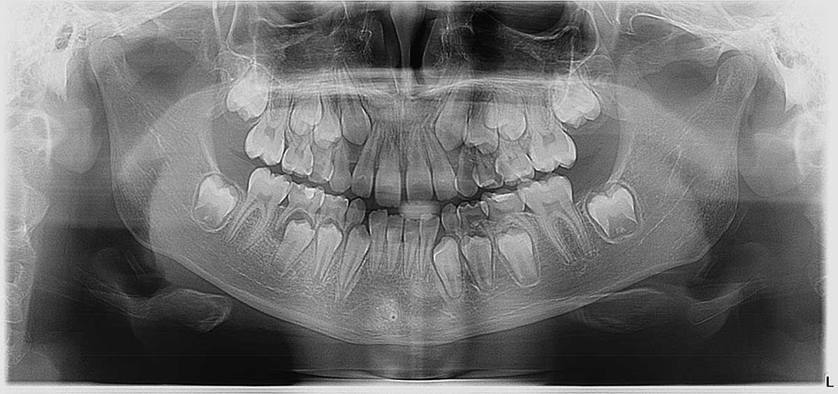
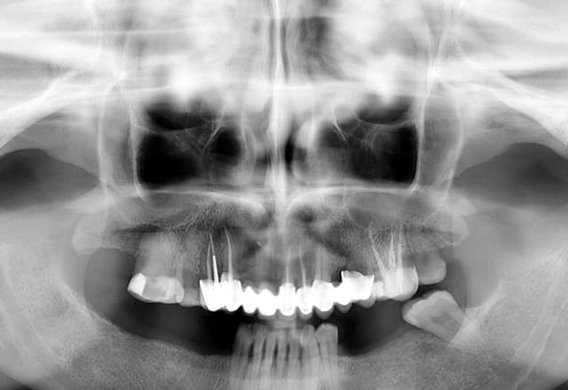
Orthopan (panoramic radiograph, digital orthopantomograph) is the most frequent image reproducing the entirety of the maxillofacial region, including the temporomandibular joints (TMJ). It can also be made in digital form on a CD (digital panoramic radiograph) to enable viewing on a computer screen. All panoramic images are made with digital CCD technology and show all the captured structures with high precision, which allows for spotting more details and making a better diagnostic application. Optimal imaging geometry creates a precise image of the teeth of the upper and lower jaw without distortion. It clearly shows pathological changes around the roots of all teeth, the state of the bone in case of periodontitis and cavity lesions on the teeth. Diagnostically speaking, it is the most important image for a timely detection of the diseases of the teeth and surrounding structures.
Orthopan shows at the same time teeth of the upper and lower jaws with surrounding tissues and structures. It is considered a basic diagnostic method in stomatology and is often developed as orientation image at the first examination. It is required in prosthetic treatment planning, in orthodontics and oral surgery when planning implant placement or removal of impacted teeth. Orthopan can be applied to the children because it is adapted small amount of radiation. The only group that is not supposed to take orthopan are pregnant women, especially in the first quarter.
Children panoramic image guarantees an excellent adjustment for children’s jaws due to the specific trajectory of its rays. Radiation is additionally decreased due to the small size of the patient. This image encompasses the entire maxillofacial complex, including the jaw joints. The possibility of front teeth overlap is reduced to a minimum. It provides for a clear view of all the details in the jaw, the positions of permanent teeth buds and possible presence of any anomalies and pathological processes.
Semi-orthopan ( half-orthopantomograph ) shows either the left or the right side of the upper and lower jaw without the shadowing made by the cervical spine. A single image can show both halves of the upper and lower jaw.
Intraoral Imaging
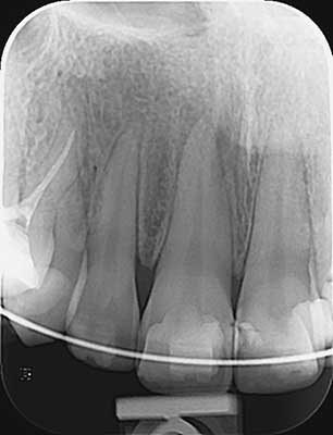
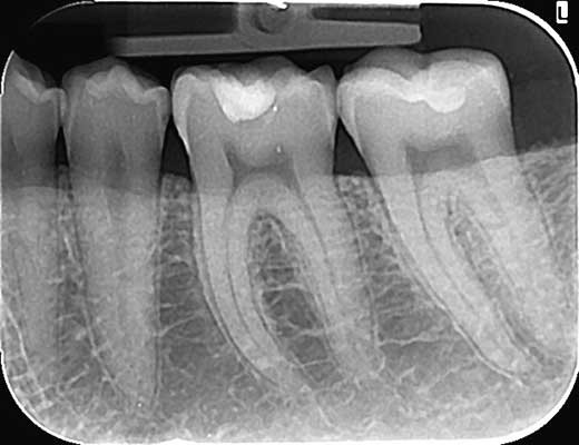
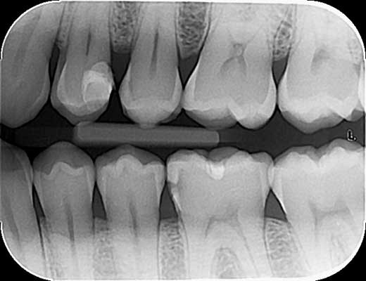
Intraoral dental images show individual teeth in real size. One image shows a single tooth or two adjoining teeth for the purpose of a regular radiographic image and the angle of the central beam.
Maxillary Sinus Imaging
Images of the maxillary sinuses. Frontal images are a tomographic section on a relatively wide section of the maxillary sinuses. They are extremely useful for implant therapy planning. Lateral images (right and left) of the maxillary sinuses are an additional projection of the frontal image of the maxillary sinus which shows the sinuses with less distortion than a standard panoramic radiograph.
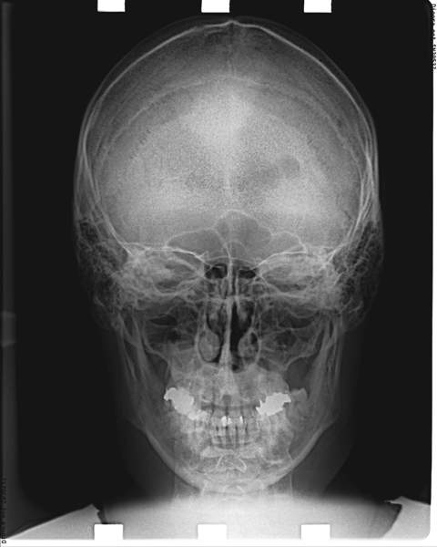
Craniogram
Craniogram (telerendgen) is an image of the face completely adapted to the patient’s morphology. It clearly shows all the bone structures and soft tissues. Craniograms can have the following projections -Latero – lateral (LL) and Posterior – lateral (PL).
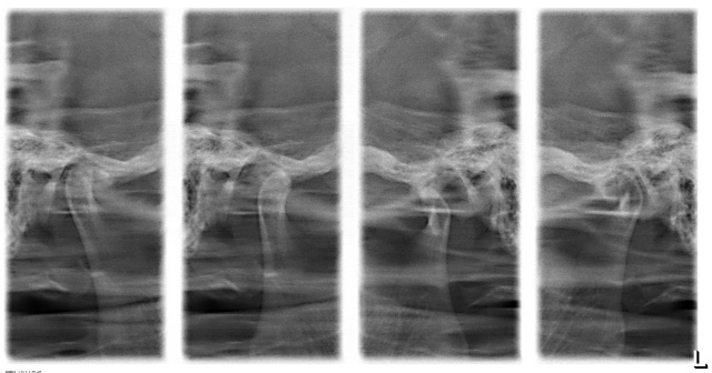
TMJ Imaging
TMJ images can be made in three ways: lateral TMJ images with the mouth closed, lateral TMJ images with the mouth open and frontal TMJ images.
We offer you the following prints on individual films:
1. frontal images and lateral images with the mouth closed (4 images: right and left frontal, right and left lateral)
2. frontal images and lateral images with the mouth open (4 images: right and left frontal, right and left lateral)
3. lateral images with the mouth closed and open (4 images)
We use cookies to ensure that we give you the best experience on our website. If you continue to use this site we will assume that you are happy with it.OkSaznaj više