These pages are for dental professionals mostly.
CBCT indications – endodontics: excellent viewing possibilities of the third dimension can be of great use in assessing periapical pathology and endodontic tooth space. Endodontic procedures can be foreseen more easily, faster and safer. With the application of dental CT scans, endodoncy gets a new diagnostic means in the assessment of:
In order to choose the appropriate implant location, precise information on the available bone and its quality, and the precise location of critical areas are needed. The location of the mandibular canal or maxillary sinus can be simply and easily determined. With a parallel view of all three dimensions, measuring functions and an implant file, it is possible to efficiently and reliably measure the implant procedure.
SINUSES – floor, position, implant direction
MANDIBULAR CANAL – direction, position, relation to roots, bone leftover for implantation
ALVEOLAR RIDGE WIDTH – quantity of cancellous and compact bone, boundary of bone and hard connective tissue
LINGUAL INCLINATION OF THE INNER MANDIBULAR SURFACE – position of the lingualis artery, direction of mandibular implant
DEPTH OF ALVEOLAR RIDGE BASE – implant direction
IMPLANT INCLINATION – toward anatomical structures, available bone, surrounding teeth
IMPLANT WIDTH – determined in relation to available bone
LOCATION OF STARTING POINT FOR BONE PREPARATION – location of implant procedure
PARA-IMPLANT RIDGE AUGMENTATION – possibility of planning augmentation before implantation
CBCT is the golden standard in implant planning, which is particularly important from the perspective of forensic medicine.
An exact 1:1 ratio enables making all the measurements necessary for determining dimensions more conclusively and precisely and for positioning the implant.
Images in the internationally standardized DICOM format can easily be transferred to most program packages for additional treatment planning. For systems of computer-guided implantology via implant templates, data on bone volume is delivered in the DICOM format.
The program contains an implant verification module, which serves to aid implant planning by rotating all images around a virtually implanted implant. This creates an enhanced view of surrounding structures. The model can be used if an implant illustration is positioned on the image. Therefore, if you wish to use this option, list the implant brand and type. We will position the implant you wish to use on the side of the image. That way you will be able to use an exact implant illustration and position it anywhere on the 3D image.
ORAL AND MAXILLOFACIAL SURGERY
The success of a surgical procedure largely depends on an accurate and precise diagnosis. Preoperative radiological diagnostics gives the surgeon a visual overview before and during the surgical procedure.
Pathological conditions and anomalies can be simply diagnosed and assessed with the help of 3D images. An unlimited number of images can be made from a single scan with a low level of radiation, making the Cone Beam CT the best device for maxillofacial surgeons.
The 3D CT offers the best diagnostic possibilities in surgical procedure planning for a vast array of surgical procedures:
EXTRACTIONS – root anatomy and position, retained roots
ALVEOTOMY – inclination, position, relation to bone and surrounding structures, phase of root development
CYSTS AND TUMORS – shape, size, spatial distribution, relation to other anatomical structures
VESTIBULOPLASTY – ridge thickness, ossification, ridge undulation
ORTHOGNATHIC SURGERY – excellent visualization and possibility of better communication between specialists, symmetry and asymmetry
RECONSTRUCTIVE SURGERY – clear view of anatomical structures, easier procedure planning, determining the middle
APICOECTOMY – periapical process, overfilling, bone defect, section behind root
FOREIGN BODIES
Foreign bodies, like roots, implants, instruments and impression or augmentation material, introduced by trauma or iatrogenic causes can be easily and precisely located. It can be determined whether the foreign bodies are located in the sinus cavity or submucously, and if any inflammation or chronic changes exist around them.
TMJ
Scanora 3D makes clear and detailed transversal, axial and sagittal images of the joint. They largely help determine intra-articular pathology and help to plan the surgical procedure on the joint. It is possible to determine the presence of degenerative processes, condylar dysplasia, joint space narrowing, eroded surfaces, subchondral sclerosis, osteophytes, cysts, trauma and primary or secondary tumors.
IMPACTED TEETH
A large number of sections and angles of the impacted tooth can be made for the purpose of an alveotomy. These images are of great help in determining the relation of molars with the bottom of the maxillary sinus or mandibular canal, the impacted cuspid with the roots of adjoining teeth and the base of the nasal cavity, mesiodens with the incisive canal, etc. It is possible to determine the presence of adjoining tooth resorption and a small-scale preparation of soft tissue can be planned. As this information is present, the specialist knows what to expect before starting the alveotomy, which reduces the possibility of potential problems to a minimum.
SYNDROMES AND MALFORMATIONS/CRANIOFACIAL DYSPLASIA
3D diagnostics gives an insight into the individual specifics and morphological characteristics of the patient. Planning and performing surgical treatment is simplified.
CLEFT LIP AND/OR PALATE
The shape and relations of unfused structures, premaxillas, the position and relation of impacted and erupted teeth, position in the ridge, relations of antagonized ridges, maxilla and mandible morphology, occlusion and the transversal relation of dental arches can all be clearly seen. Symmetry or the rate of asymmetry of the septum, dental arches and palate can be determined. CBCT enables a clear 3D anatomical image, the assessment of cleft type and size and an assessment of residual defects after osteoplasty. Determining the clinical status has become simpler, and therapy planning more efficient.
OROANTRAL COMMUNICATION
Precise diagnostics of the root-sinus relation prevents the formation of an oroantral communication, while determining its position, size and shape facilitates therapy. Diagnostics of the root-sinus relation enables prevention, and determining the position, size and shape facilitates therapy.
MAXILLARY SINUSES
Symmetry, shape, distribution and relation to adjoining structures are easily determined with 3D diagnostics. The presence of inflammations, foreign bodies, cysts or tumors can be better diagnosed. Planning operative procedures is faster and simpler. It is simpler to identify the positions of retained roots, the need for maxillary sinus floor augmentation and membranes, while sinus lifting can be more easily planned.
TRAUMA AND FRACTURES
Trauma of the maxillofacial region usually contains combined injuries of different structures. By using 3D diagnostics, it is possible to obtain a large quantity of important information during the primary phase of trauma diagnostics, since the image can encapsulate the entire region. With all potential or determined viscerocranium fractures, it is necessary to complement 2D diagnostics with a 3D CT.
The ability of the 3D CT to make precise measurements and axial images can be used to view the characteristics of airways and diagnose apnea, pharyngeal diseases and diseases of the upper respiratory tract.
A sagittal view of the upper respiratory tract or nasal section of the pharynx can clearly show the relation of adenoid tissue (so called third tonsil) with the posterior section of the nasal concha.
A transversal view helps diagnose the condition of the airways. A view of the posterior section of the pharynx is used in determining anatomical obstacles made by hypertrophic adenoid tissue. Air passage can be measured in three dimensions.
When planning orthodontic therapy for different anomalies, a 3D visualization of jaw anatomy can be of great use. It achieves an excellent view of the position and inclination of the teeth and roots, the location and direction of impacted teeth and the relation of the root and the cortical bone. Anatomical structures such as the incisive canal, TMJ, the angle of the mandible, the retromolar space, and general maxilla-mandible relations are much more visible, which provides for an application of 3D diagnostics in planning orthodontic and orthognathic surgery therapy. Apart from that, digital models of the entire jaw can be permanently stored on CDs, which decreases the need for space taken by study or control gypsum casts.
Tooth position
It is possible to determine the inclination and position of roots, periodontal thickness and space, tooth axis and the rotations of individual teeth. In the planning phase, we can accurately identify the equator of every tooth and precisely position the brackets with regards to the desired position. We decrease the need for rebonding and additional bracket positioning. The straight-wire concept reaches its full meaning through accurate planning and root position visualization.
Impacted teeth
With the help of 3D CT devices it is now possible to precisely determine the location of impacted cuspids and molars, their direction, position and relation to adjoining teeth. It is also possible to determine whether resorption of the roots of adjoining teeth has occurred and to what extent. Planning a combined orthodontic-surgical therapy is largely facilitated. Moving the impacted tooth after surgical visualization can be foreseen more precisely and the total orthodontic therapy time can be shortened.
Maxillary sinus
Establishing the relation between the back teeth of the upper jaw and the cortical bone of the sinus floor is important for planning tooth shift.
Incisive canal
When planning a diastema closure or a front teeth torque, it is important to determine the size, shape and position of the incisive canal, since it can block an orthodontic shift.
Arch expansion
Assessments on whether there is enough space in the alveolar bone to perform a tooth arch expansion and whether the patient’s anatomy requires a forced palate expansion can be made more precisely with the help of 3D images.
Cortical bone and tooth eruption disorders
Measuring the size of the alveolar bone is a decisive factor in determining the potential of an orthodontic shift. It is possible that structures exist within the alveolar bone, e.g. corticalization, which would stop tooth shifting during orthodontic treatment. Distances of individual roots from the cortical bone are also easily determined.
Orthognathic pre-surgical treatment and cleft treatment
Planning a combined orthodontic-surgical therapy with 3D diagnostics can significantly speed up the treatment course, enhance communication between specialists and enable a more precise planning of all treatment procedures.
TMJ
It is possible to observe the anatomy of the condyle, articular fossa, atricular eminence, condyle position based on intercuspidation and condyle symmetry.
Retromolar space
3D diagnostics fully enables molar and retromolar space assessment, which is a decisive factor in extracting wisdom teeth or any other teeth in the molar region.
3D cephalometric analyses
3D images enable us to precisely locate cephalometric landmarks and get additional insight into the patient’s craniofacial characteristics.
In periodontal practice, the 3D CT is used for a more detailed examination of the spatial relations of periodontal pockets, furcation openings and the seriousness of bone recession.
PERIODONTAL POCKETS
FURCATIONS
RECESSIONS
CBCT Indications
Cone Beam CT has a series of advantages when compared to conventional radiograms. Due to its low levels of radiation and high diagnostic value, the CBCT can be applied in almost all branches of dental medicine.
Scientific work
3D CT offers excellent possibilities for diverse types of scientific research. Seeing that images are standardized and that calibration is performed regularly, the images offer completely accurate and precise data ready for further processing. Very detailed morphometric data can be easily and simply run multiple times with perfect accuracy.
Apart from biometrics, it is possible to study human diversity, hard tissue structure, their density, shape and variability, anatomical characteristics, pathological changes, anomalies and malformations, etc. CBCT can be applied in various areas from anatomy and anthropology, physiology, histology, densitometry, pathology to case studies, and studies of nervous system biomechanics and occlusal relationships.
High quality images with a 3D view and measurements can be produced for the purposes of scientific publications. Case studies of rare medical cases can be permanently stored in a 3D format and later used for additional research.
ANTHROPOLOGY
ANATOMY AND PATHOLOGY
DENSITOMETRY
MORPHOMETRICS
OCCLUSION
SYNDROMES
CASE STUDIES
IMAGES FOR PUBLICATION
Download and install DfS
Once you have opened the program:
– Click on Patient list then click Show (F3) – a window will open at the bottom of the screen showing your patients and several examples. We recommend you always keep this window open to keep an eye on everything more easily.
– Open Cranex D. Double click on the panoramic radiograph in the top right corner. An active panoramic radiograph image will appear with an Image Toolbox on its right. If you right-click on the small image on Image Toolbox, a window showing all the options shown on Image Toolbox will pop up. Select all the options.
If you are not satisfied with the view on your monitor, adjust it by opening Options – General setup, selecting the Enhance tab and customizing Matrix size in the white box. You can only insert odd values from 1 to 49 (e.g. 11, 23, 41).
Note: If you are having difficulties downloading DIGORA FOR SCANORA, do not hesitate to send us an e-mail describing the problem on help@testa.lauc.hr.”>help@testa.lauc.hr
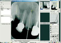
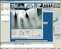
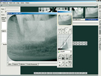
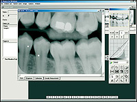
Digora for Scanora
DIGORA for Scanora has all the tools necessary to view and process digital images, and precisely measure and enhance the viewability of certain regions of panoramic radiographs, craniograms or dental photographs. “Implant Module Library”, a built-in module for implant planning, enables a precise planning of positioning different implants on a digital panoramic radiograph. An extended implant data base includes new models of leading global manufacturers and facilitates your choice of implants as it does their positioning on all dental images.