Digital radiographic images in dental practice
Dental therapy diagnostics and planning largely rely on technological advances and the development of medical equipment. The aim of perfecting medical equipment in radiographic diagnostics is showing the so called anatomical reality, i.e. achieving an image of the anatomical structure that is as accurate as possible. Radiographic images of the teeth and jaws are the most frequent diagnostic tool that helps dentists of all branches and specialties identify anatomical and pathological occurrences, as well as measure the sizes of the structures of the craniofacial system and determining their form.
Advantages of digital images
Radiographic images contain information and they can be made by means of conventional analogue procedures, or by means of more advanced digital procedures. Digital images simplify handling, they can be faithfully rendered on computer screens and later used on the computer for all necessary procedures. It is possible to make a software analysis of bone density, zoom in on individual details, change contrast and color desired structures. Digital storing is made easier by the fact that a large number of images can be stored on hard disks or portable media and sent online. But most importantly, biologically speaking, digital imaging technology has enabled a 30-98% decrease in radiation doses delivered to the patient.
Means of storing information
During digital imaging, CCD sensors turn the electromagnetic energy of x-ray beams into an electric impulse. Electric impulses have different photosensory values and a digitalization board turns them into pixels, which are assembled into horizontal raster lines. The raster lines make up a pixel matrix called a map. Each pixel, i.e. each picture unit, has its dimension and intensity, which determines the surface area and degree on the grey-scale representing tissue structure on the radiographic image.
Application of digital images
The advantage of digital radiography is seen in the fact that, by applying different algorithms, a digital image can be enlarged, sharpened, made more grainy, made less intense (“smoothed out”) and its specific parts can be isolated. However, the quantity of stored information does not increase when applying the processing procedures. This is why the image must be thoroughly prepared in a radiographic laboratory by means of specialized software which will make it possible to additionally process the image by increasing its total information count (its pixel diversity) before storing.
Equipment


The most advanced member of the renowned CRANEX™ product family is now available.
The new CRANEX™ 3Dx unit extends possible application areas of 2D panoramic and cephalometric imaging to accurate 3D imaging from a single tooth to the entire cranio-maxillofacial region. Two optional large fields-of-view are perfect for diagnostic tasks in the ENT areas. The icing on the cake is the low dose imaging programs with excellent diagnostic values and the Automatic Exposure Setting (AES) in 2D panoramic and cephalometric imaging, as well as in 3D – a world first AES hybrid.
CRANEX ™ 3Dx spans four decades of experience in designing imaging solutions and offers a real diagnostic value.
Top performance for top performers
- Excellent image quality in 2D and 3D needed for accurate diagnosis
- You can choose exactly the right FOV size according to ROI and diagnostic task; no need to over-radiate the patient just to make sure you have the ROI in the image
- Thanks to the SOREDEX® MiniDose solution, you can take advantage of 3D diagnostic data even in dose sensitive cases like children, or reduce the radiation load for the patient during treatment follow-up
- User-definable resolution settings help you to choose just the right dose following the ALARA principle
Ease of use means time-savings in your daily workflow
- ClearTouch™ control panel with simplified selections is located conveniently in the unit column for quick access
- The renowned SOREDEX™ patient positioning system helps your patient to keep still during exposure, which is one of the main prerequisites for good image quality
- The Automatic Exposure Setting (AES) function suggests exposure values based on patient size. Now, as a world first, CRANEX™ 3Dx offers this valuable feature for 3D imaging too.
Versatile options for optimized imaging
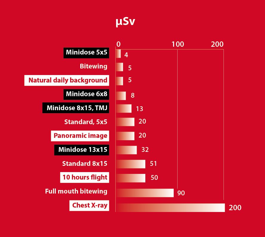
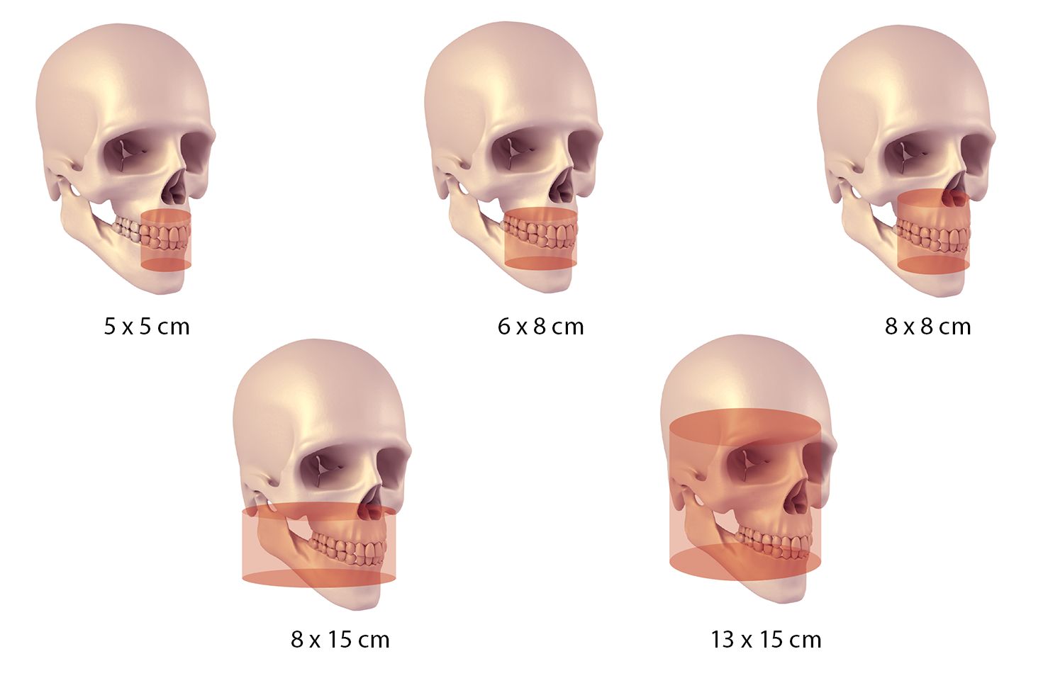
- 5 fields of view accommodate multiple application areas in the head and neck region: 5 x 5 cm, 6 x 8 cm, 8 x 8 cm as standard and optional 8 x 15 cm and 13 x 15cm
- The Minidose™ solution (MDS) enables very low dose imaging, e.g. in 5 x 5 cm FOV, effective dose is only 1/5 of a panoramic image! Good image quality can be maintained thanks to SARA™ technology
Intraoral diagnostics. In a flash.
5th generation DIGORA™ Optime unit continues the intraoral imaging revolution started by the original DIGORA® 20 years ago. The concise, easy to use design makes intraoral imaging more intuitive and efficient than ever before. Uncompromised image quality, with literally no buttons pressed.
Smart
- Delivers always images with good contrast with its Auto-Optimization –feature
- Smart automated functions and economical network sharing
- Latest generation, wireless and durable imaging plates with patented IDOT™ identification system (optional)
- Comfortable and safe with unique hygiene accessories
Features. Repeatable image quality
- Fully automatic and optimized image processing
- Exposure assistant –feature guides for correct exposure settings
- High quality images are displayed in just seconds
- Processing speed is not dependent on image resolution
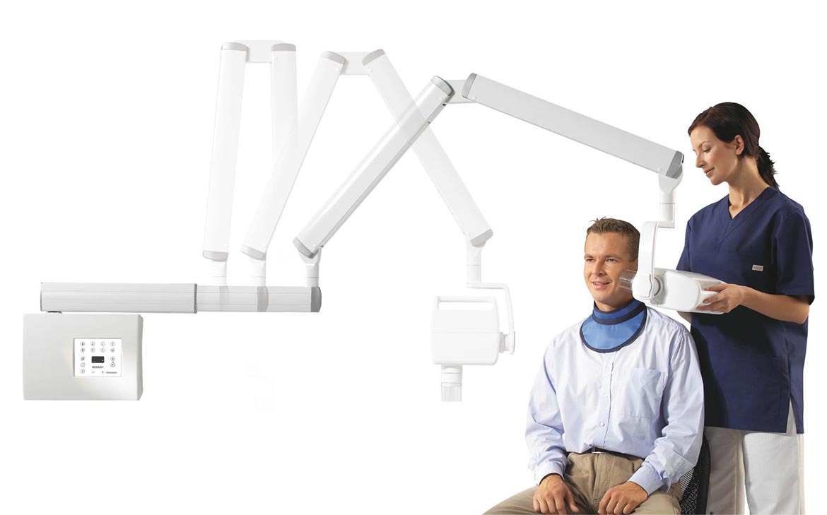
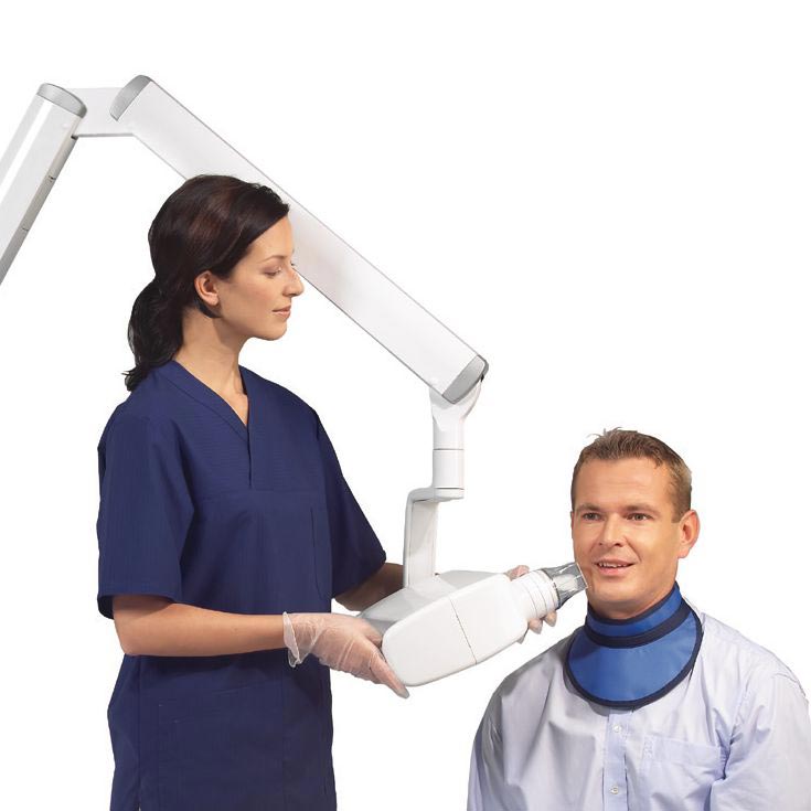
Amazing images. At an arm’s reach.
MINRAY™ unit is a compact, versatile intraoral X-ray, with a high frequency DC output for minimized soft radiation. Designed for efficient workflow, MINRAY™ features a unique, adjustable horizontal arm, steady arm system and a special tubehead design dedicated to the comfort of both the patient and the user. Advanced intraoral imaging, easily at your hands.
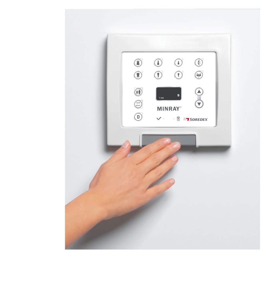
Easy
- User-friendly control panel in the wall box
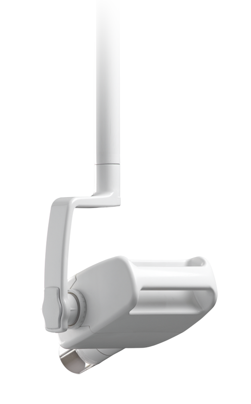
- Functionally designed tubehead handle allows one-handed movement
- Tubehead handle has no holes or gaps which are difficult to keep clean
- Remote control panel (optional) with large push bar exposure switch, can be mounted in almost any location
Precise
- Precisely repeatable time and kV enable superior image quality
- The Very High Frequency (VHF) constant potential (DC) technology improves image quality and is not affected by line voltage fluctuations
- Steady arm system ensures smooth operation and stable exposure
- Strong mechanical construction manifest long service life
Adjustable
- Unique extendable horizontal arm design for optimized reach and ergonomics
- Arm length is adjusted and fixed at the time of installation and it can be readjusted later, if MINRAY™ unit needs to be re-located
- Eight preprogrammed settings or easily store your own in the unit memory
- Variety of beam limiting cones
- Remote control panel (optional)
- Unit mount cable kit (optional)
- Mobile stand (optional) allow one MINRAY™ x-ray unit to be shared in multiple operatory practices
Clear and easy controls
- User-friendly control panel
- Installed in a wall box or remote mounted in almost any location
- Dual installation: one in the wall box and one outside the operatory
- 8 pre-programmed settings
- Exposure time range 0.02 and 3.2
- Choice of 60 and 70 kV for optimized contrast
- User defined settings stored easily
Aim for the highest diagnostic performance
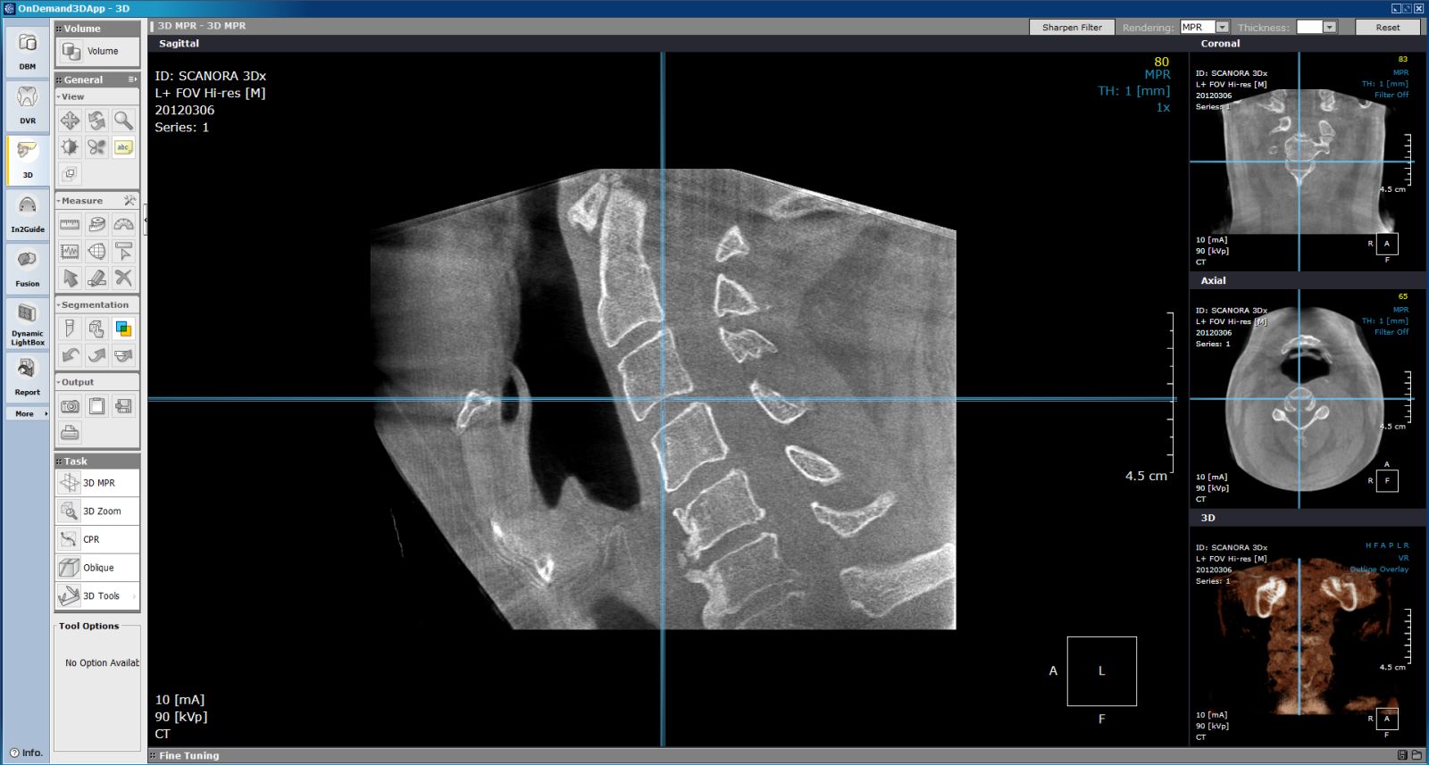
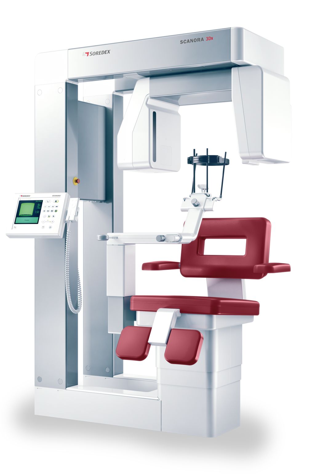
SCANORA™ 3Dx is a cone beam CT imaging device aimed for contemporary practices that offer multi-specialty treatment services. It helps ENT (ORL) and dento-maxillofacial surgeons to make accurate diagnostics, precise treatment planning for implant, ENT and oral surgery, and efficiently follow up on the treatment.
We’ve built SCANORA™ 3Dx unit with the patient in mind, managing to balance excellent image quality with low radiation doses. The design supports convenient patient positioning and easy workflow. Compared to the sister unit SCANORA™ 3D, the 3Dx offers a greater variety of field-of-view options, from small highlight scans to large, overall head and neck imaging.
Flexible
- Variety of software options available from Soredex
- Optional CCD – RealPAN™ sensor for high quality dental panoramic imaging, with AutoSwitch™ 2D/3D mode change (no manual sensor change)
Selection of FOV’s for variety of diagnostic tasks
- Six, plus two optional, fields-of-view from 50 x 50 mm to 240 x 165 mm
- Standard and high resolution available for optimizing the protocol according to diagnostic task (ALARA principle)
Low-dose 3D imaging made easy
- Cone beam technique
- FOV and resolution freely selectable according to region of interest (ROI)
- Adjustable mA
- Innovative reconstruction algorithm SARA™
- Pulsed x-ray generation